
Fig. 1
- ID
- ZDB-IMAGE-080506-23
- Genes
- Publication
- Chuang et al., 2001 - Zebrafish genes rx1 and rx2 help define the region of forebrain that gives rise to retina
- All Figures
- Figures for Chuang et al., 2001
|
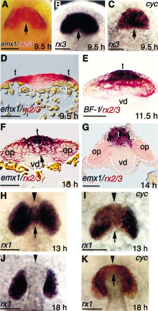
Fig. 1 Expression of rx genes during neurulation in wild type and cyclops embryos. (A, B) Normal expression pattern of rx in the rostral neural plate (A) in relation to emx1 expression or (B) by itself. (C) Expression of rx3 in cyclopsb16 embryos at the neural plate stage. Note the absence of the caudal indentation at the midline [compare arrow in (C) with arrows in (A) and (B)]. (D-G) Transverse sections of whole-mount, double in situ preparations at the level of (D) the retinal field or (E-G) the optic primordia. Note the relationship between retinal precursors (marked by rx, in red) and telencephalic precursors (marked by emx1 or BF-1, in purple). The unlabeled cells in the ventral neural keel are ventral diencephalic precursors. (H-K) Comparison of rx1 expression in wild type (H, J) and cyclops embryos (I, K). The arrows indicate the suppression of rx1 expression at the caudal midline. The arrowheads indicate the region in which rx1 expression differs between wild type and cyclops embryos. Note that rx1 is not expressed in the rostral hypothalamus in wild type embryos, as reflected by an indentation at the midline on the rostral side (arrowhead). In cyclops embryos (I, K), the rostral hypothalamus is missing, and the two eyes are fused in the rostral midline (arrowhead). Rostral is up in (A-C) and (H-K). op, optic primordium; t, telencephalon; vd, ventral diencephalon. Scale bars: 100 μm.
Reprinted from Developmental Biology, 231, Chuang, J.-C. and Raymond, P.A., Zebrafish genes rx1 and rx2 help define the region of forebrain that gives rise to retina, 13-30, Copyright (2001) with permission from Elsevier. Full text @ Dev. Biol.