
|
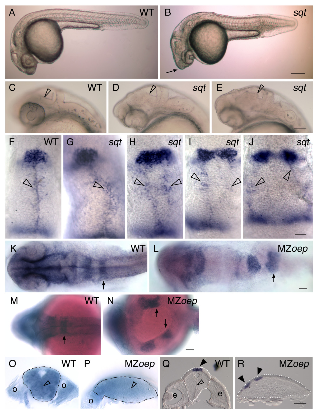
Fig. 3 Correlation between an expanded or divided pineal and an open neural tube. (A-E) Lateral views of live embryos at 1 dpf, anterior to the left. (A) While the head of the WT embryo is smooth and rounded, (B) the head of the sqt embryo is pointed (arrow). (C-E) Higher magnification of the anterior embryo reveals variability in the brain morphology of sqt mutants. In (C) WT embryos and (D) some sqt mutants, the border between the tectum and tegmentum (open arrowheads) appears as a smooth, straight line. (E) However, in some sqt mutants the border appears to be abnormally shaped or indistinct (open arrowhead), suggesting that the morphology of tectum or tegmentum is perturbed. (F-J) Embryos were fixed at 1 dpf, and processed for in situ hybridization with antisense probes for the pineal gene otx5 and the dorsal neural tube gene wnt1. In (F) WT and (G) sqt embryos with a single, round pineal anlage, the wnt1 expressing cells (open arrowheads) form a single domain along the dorsal neural tube. In contrast, sqt embryos with an (H) elongated or (I-J) divided pineal anlage have two parallel lines of wnt1 expressing cells. (K-P) Embryos were fixed at 1 dpf, processed for in situ hybridization with an antisense probe for epha4a, and then either (K-N) imaged in dorsal view, anterior to the left or (O, P) cut through epha4a-expressing rhombomere 5 to bisect the embryo into anterior and posterior halves. The locations of the otic vesicles (o), rhombomere 5 (arrows), and midline (open arrowhead) are indicated. A potential region of midline is marked by the open arrowhead in P. (Q, R) 14 μM frozen cross sections through the diencephalon of 1 dpf (Q) WT or (R) MZoep embryos stained for otx5 expression. The midline of the brain (open arrowhead), and pineal precursors (closed arrowheads) are indicated. Dotted lines outline the neural tubes in panels O-R. Scale bars: 200 μm (A,B), 100 μm (C-E), 30 μm (F-J), 50 μm (K-R).