
|
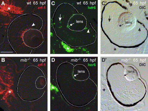
Fig. 3 Müller glia differentiation is inhibited in mib-/- retinas at 65 hpf. Müller glia in the retina (outlined) expressed zrf-1/GFAP (arrowhead) as did astrocytes and/or radial glia in the brain (*) of wild-type embryos (A). In contrast, zrf-1/GFAP expression was absent in the retina (outlined) but present in the brain (*) of mib-/- embryos (B; N = 10). (C, D) Expression of her6/hes1 in wild-type and mib-/- 65 retinas. (C′, D′) Differential interference contrast (DIC) images of the retinas in (C and D; N = 8 embryos). (C, C′) In the wild-type embryos, her6/hes1 expression was present in the brain (*) and in the germinal zone (arrowhead), lens epithelium (small arrow) and inner nuclear layer (large arrow) of the retina. (D, D′) The mib-/- embryos expressed her6/hes1 in the brain (*) but expression in the eye was restricted to the lens epithelium (small arrow). Scale bar = 50 μm (A–D).
Reprinted from Developmental Biology, 278(2), Bernardos, R.L., Lentz, S.I., Wolfe, M.S., and Raymond, P.A., Notch-Delta signaling is required for spatial patterning and Muller glia differentiation in the zebrafish retina, 381-395, Copyright (2005) with permission from Elsevier. Full text @ Dev. Biol.