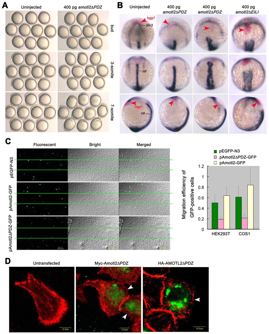
Fig. 6 Effect of amotl2 mutants on cell migration and structures. (A) Effect of overexpression of amotl2ΔPDZ mutant on cell movements of zebrafish embryos. Embryos were injected at the one-cell stage and observed at the indicated stages. Embryos were managed to position with animal pole or anterior to the top. (B) Expression of hgg1 (red), dlx3 (blue) and ntl (blue) at the two-somite stage. Top panel, anterodorsal views; middle panel, dorsal views with anterior to the top; bottom panel, lateral views with anterior to the top and dorsal to the right. Injection of amotl2ΔPDZ mRNA led to varying degrees of defects in convergent extension (in the second and third columns). Injection of amotl2ΔEILI mRNA also caused defective convergent extension (fourth column). The hgg1 expression domain is indicated by arrowheads. (C) Expression of Amotl2ΔPDZ-GFP or Amotl2-GFP inhibited or promoted cell migration in vitro, respectively. Wound-healing assays were done both in HEK293T (shown on the left) and in COS1 cells. The wound area was between two lines. The bar graph at the right shows statistical data from three experiments with standard deviations. The migration efficiency of GFP-positive cells was calculated as percentage of GFP-positive cells in the wound-healing area/percentage of GFP-positive cells in the non-wound area. (D) F-actin distribution was abnormal in HeLa cells transfected with either Myc-Amotl2ΔPDZ (derived from fish) or HA-AMOTL2ΔPDZ (derived from human). Cells were stained with phalloidin 24 hours after transfection. Arrowheads indicate Amotl2-expressing cells. Scale bars: 10 μm.
Image
Figure Caption
Figure Data
Acknowledgments
This image is the copyrighted work of the attributed author or publisher, and
ZFIN has permission only to display this image to its users.
Additional permissions should be obtained from the applicable author or publisher of the image.
Full text @ Development

