
Fig. 4
|
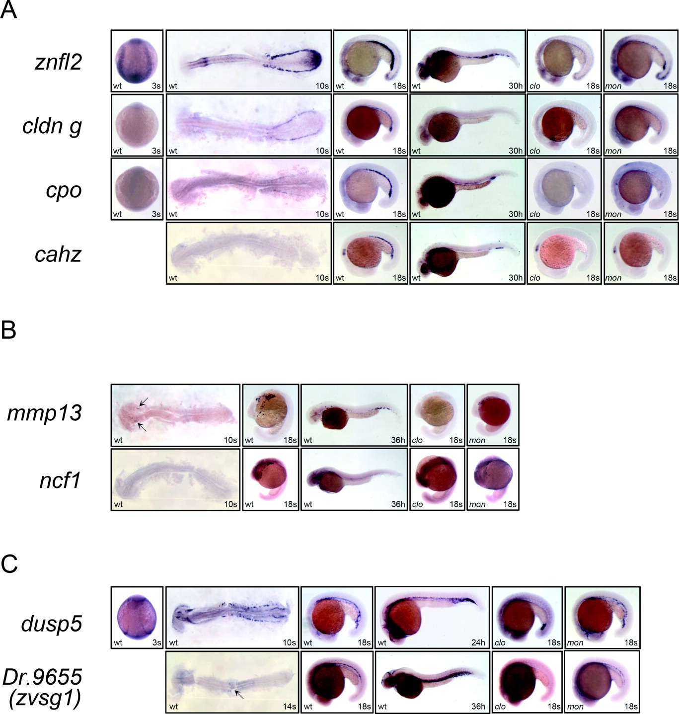
Fig. 4 Temporal and spatial expression of znfl2, cldn g, cpo, cahz, mmp13, ncf1, dusp5, and zvsg1. A: Whole-mount in situ hybridization shows that znfl2 (top panels), cldn g (second panels), cpo (third panels), and cahz (bottom panels) are expressed in erythrocytes. B: Whole-mount in situ hybridization indicates that mmp13 (upper panels) and ncf1 (lower panels) stain myeloid lineage cells. C: Whole-mount in situ hybridization shows that dusp5 (upper panels) and zvsg1 (lower panels) express in the head and trunk vessels. The 3-, 10- and 14-somite stage embryos are dorsal view (3s) and flat-mount in dorsal view (10s and 14s), whereas rests of the embryos are lateral view. In all the panels, the 3-somite stage embryos are oriented anterior to the top and the rest are oriented anterior to the left.