
Fig. 6
|
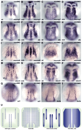
Fig. 6 Her3 and Her9 are required for formation of the inter-proneuronal domains. (A-P) Expression of neurog1 (A-D), deltaA (dla; E-H), neurod4 (I-L) and elavl3 (M-P) in wild-type embryos that received 2 ng control MO (A,E,I,M) or 1 ng her3-MO1 and 2 ng her9ATG-MO (C,G,K,O), or in mib mutant embryos that received control MO (B,F,J,N) or her3/her9-MO (D,H,L,P) at the one-somite stage. Dorsal views. (Q-T) Expression of islet1 (isl1, a marker for primary motoneurons and RB neurons) and tlx3a (a marker for RB neurons) in the control (Q,S) and the her3/her9-MO-injected embryos (R,T) at the three-somite stage. Expression of islet1 and tlx3a was not affected in the her3/her9 morphant embryos. (U) Schematic representation of the expression profiles of neurog1, dla, neurod4 and elavl3 in wild-type or mib embryos that received control or her3/her9-MO injections. The expression of these genes became ubiquitous in the neural plate, except for the midline region including the floor plate in the her3/her9 morphant embryos, and became homogenous within the proneuronal domains in the mib mutant embryos. The her3/her9 morphant mib mutant embryos showed ubiquitous and homogeneous expression of these genes in the neural plate, except for the midline region.