
Fig. 9
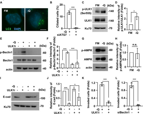
Glutamine deprivation enhances autophagy to promote primary ciliation and cell invasion. A Autophagy is activated by Gln deprivation. Autophagy was observed by immunofluorescence staining with antibodies against LC3 in PANC-1 cells cultured in full-medium (FM) or Gln-deprived medium (-Q). DNA were stained with DAPI. Scale bar: 10 μm. B Depletion of ATG7 inhibited primary cilia under Gln deprivation in PANC-1 cells. Quantitative results of ciliated cells in the absence or presence of siRNA against ATG7. C-D Gln deprivation activated ULK1 in PANC-1 cells. C Extracts of PANC-1 cells cultured in full-medium (FM) or Gln-deprived (-Q) medium were analyzed by Western blotting with antibodies against phosphorylated ULK1 (p-ULK1), ULK1, phosphorylated AMPK (p-AMPK), AMPK, Ku70, and actin. D Quantitative results for the relative intensity of p-ULK1/ULK1 in C. E-F Inhibition of ULK1 alleviated Beclin1 phosphorylation under Gln deprivation. (E) Extracts of PANC-1 cells were analyzed by Western blotting with antibodies against phosphorylated Beclin1 (p-Beclin1), Beclin1, E-cadherin (E-cad), Ku70, and actin. F Quantitative results for the relative intensity of p-Beclin1/Beclin1 in E. (G-H) AMPK was not activated during Gln deprivation in PANC-1 cells. G Extracts of PANC-1 cells cultured in full-medium (FM) or Gln-deprived (-Q) medium were analyzed by Western blotting with antibodies against phosphorylated AMPK (p-AMPK), AMPK, and Ku70. H Quantitative results for the relative intensity of p-AMPK/AMPK in G. (I-J) Inhibition of ULK1 alleviated EMT under Gln deprivation. I Extracts of PANC-1 cells were analyzed by Western blotting with antibodies against E-cadherin (E-cad) and Ku70. J Quantitative results for the relative intensity of E-cad in I. K-L Inhibition of ULK1 or depletion of Beclin1 alleviated PANC-1 cell invasion under Gln deprivation. Quantitative results of invaded cells in the absence or presence of (K) ULK1 inhibitor (ULKi) or (L) siRNA against Beclin1 (siBeclin1). ***:p < 0.001. These results are mean ± SD from three independent experiments |