
Fig. 2
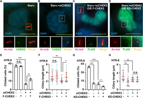
CHEK2 regulates primary cilia via its kinase activity. A-D CHEK2 is localized at the base of primary cilia. HTR-8 cells were serum-starved for 24 h and then examined for (A–B) endogenous CHEK2 under (A) control (Starv.), B CHEK2-deficient (siCHEK2), exogenous (C) FLAG-tagged wild-type CHEK2 (O/E F-CHEK2), or (D) kinase-dead CHEK2 (O/E KD-CHEK2) in CHEK2-deficient cells. Signals were detected by immunofluorescence staining with antibodies against acetylated tubulin (Ac-tub), CHEK2, and FLAG. DNA were stained with DAPI. Scale bar: 10 μm. E-F Overexpression of wild-type CHEK2 rescued the defect of (E) ciliated cells and (F) the length of primary cilia in scramble control or CHEK2-deficient (siCHEK2) HTR-8 cells with or without overexpressing FLAG-tagged CHEK2 (F-CHEK2). G-H Overexpression of kinase-dead CHEK2 inhibited primary cilia. Quantitative results of (G) ciliated cells and (H) the length of primary cilia in scramble control or CHEK2-deficient (siCHEK2) HTR-8 cells with or without overexpressing kinase-dead CHEK2 (KD-CHEK2). n.s.: no significance, *p < 0.05, **p < 0.01, ***p < 0.001. These results are mean ± SD from three independent experiments |