|
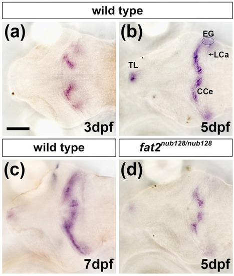
Expression of fat2 in wild‐type and mutant zebrafish larvae. (a–d) Whole‐mount in situ hybridization showing expression of fat2 mRNA at (a) 3 dpf, (b) 5 dpf, and (c) 7 dpf in wild‐type larvae, and (d) at 5 dpf in fat2nub128/nub128 mutant larvae, detected using a digoxigenin‐labeled antisense probe (n = 5). Dorsal views with anterior to the left. In wild‐type larvae, signal is detected in cerebellar granule cells, including the CCe, EG, and LCa. In mutant larvae, signal is reduced in the CCe and nearly absent in the EG and LCa. Scale bar: 100 μm in (a) (applies to a–d). Images are representative of the samples examined, and the observed expression patterns were consistent across all samples (n = 5).
|