
FIGURE 4
- ID
- ZDB-FIG-260626-15
- Publication
- Rezi et al., 2026 - ZDHHC5 interacts physically and functionally with DLG1 at primary cilia and regulates ciliary length and kidney morphology
- Other Figures
- All Figure Page
- Back to All Figure Page
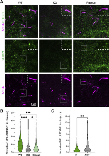
STXBP1 localizes to the base of primary cilia in a DLG1-dependent manner. (A) IFM analysis shows ciliary base localization of STXBP1 in control (WT) IMCD3 cells, which is lost in Dlg1 KO cells, and restored upon stable expression of mCherry-DLG1 in the KO line (Rescue). Cells were serum-starved for 24 h and subjected to IFM analysis using antibodies against STXBP1 (green) and acetylated α-tubulin (AcTub; magenta), which stains the ciliary axoneme. Insets show zoom-ins of the cilium-basal body axis. (B) Quantification of MFI of STXBP1 at the ciliary base of the indicated cell lines, based on images as in A (N = 3; WT n = 143, KO n = 107, Rescue n = 98). (C) Quantification of MFI of STXBP1 along the cilia of the indicated cell lines, based on images as in |