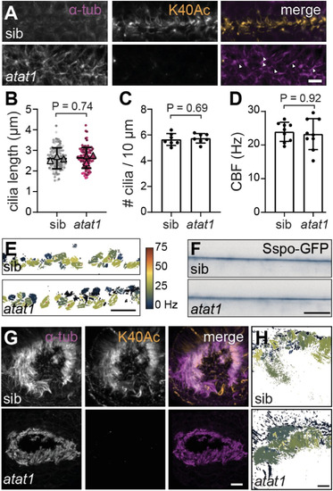
⍺TAT1 is required for ⍺-tubK40Ac in cilia, but loss of ⍺TAT1 does not impair cilia function (A) Immunofluorescence of the central canal in 24 hpf wild-type sibling and atat1 mutant embryos stained for ⍺-tubK40Ac and ⍺-tub. Arrowheads indicate cilia. Scale bar, 10 μm. (B) Superplots of central canal cilia length. Dots represent individual cilia; triangles indicate per-fish means (n = 3 per genotype). Bars show mean ± standard deviation. P = 0.74 (unpaired t-test). (C) Central canal cilia density, defined as the number of cilia per 10 μm. Bars show mean ± standard deviation. P = 0.69 (unpaired t-test). (D) Central canal cilia beat frequency (CBF). Bars show mean ± standard deviation. P = 0.92 (Mann-Whitney U test). (E) Heatmap of CBF measurements shown in (D). Scale bar, 10 μm. (F) Representative frame from live imaging of Sspo-GFP, which labels the RF, in the central canal. Scale bar, 10 μm. (G) Immunofluorescence of the olfactory placode at 6 dpf in wild-type sibling and atat1 mutant embryos stained for ⍺-tubK40Ac and ⍺-tub. Scale bar, 5 μm. (H) Heatmap of olfactory placode CBF measurements, with the key as given in (E). Scale bar, 5 μm.
|
