
Figure 3
- ID
- ZDB-FIG-260530-203
- Publication
- Mencacci et al., 2026 - Pathogenic variants in BORCS5 Cause a Spectrum of Neurodevelopmental and Neurodegenerative Disorders with Lysosomal Dysfunction
- Other Figures
- All Figure Page
- Back to All Figure Page
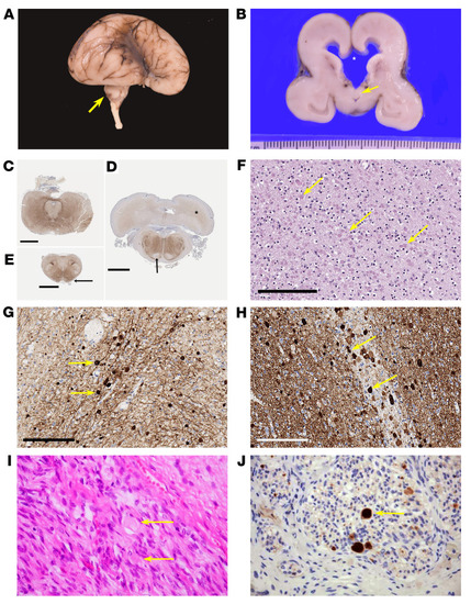
Pathological characterization of cases with bi-allelic LoF BORCS5 variants. (A) Brain of case F-VII:1 demonstrating hypoplastic temporal lobes; a delayed, smooth Sylvian fissure; and a markedly hypoplastic cerebellum (arrow). (B) Coronal section, demonstrating ventriculomegaly. The corpus callosum is reduced to a thin membrane and has ruptured (indicated at *). The septum is ruptured, and the fornices (arrow) are descended and lie on the roof of the third ventricle. (C–E) Immunohistochemistry of neurofilament light chain in whole mounts of posterior fossa structures showing midbrain with minute aqueduct and hypoplastic cerebral peduncles (C); the caudal pons with very small and smooth inferior olivary nuclei, hypoplastic middle cerebellar peduncles, absent corticofugal tracts (arrow) and poorly defined dentate nuclei (*) (D); absent pyramids (arrows) and inferior cerebellar peduncles in the medulla (E). Scale bars: 3 mm (C–E). (F–H) Histological analysis of the same patient, with H&E staining and immunohistochemistry demonstrating innumerable pale eosinophilic axonal spheroids (arrows) in the internal capsule (F); strong positive staining of the axonal spheroids for neurofilament light chain (G) and α-synuclein (H). Scale bars: 200 μm (F–H). (I and J) Peripheral nerves of individual F-V:2 demonstrated numerous axonal spheroids (arrows) (I), which stained strongly positive for β-amyloid precursor protein (J). |