
Figure 2
- ID
- ZDB-FIG-260530-202
- Publication
- Mencacci et al., 2026 - Pathogenic variants in BORCS5 Cause a Spectrum of Neurodevelopmental and Neurodegenerative Disorders with Lysosomal Dysfunction
- Other Figures
- All Figure Page
- Back to All Figure Page
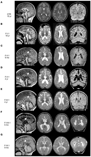
Neuroimaging findings in individuals with BORCS5 missense and splice-site variants. Brain MRI studies of a control individual performed at 15 years of age for comparison (A) and of individuals F-I:1 (B), F-II:1 (C and D), F-III:1 (E), F-VIII:1 (F), and F-IX:1 (G). Presented are sagittal T1-weighted images (first column), axial T2-weighted images (second and third column), and coronal FLAIR or T1/T2-weighted images (last column). There is moderate to severe cerebral atrophy and loss of white matter volume with consequent ventricular dilatation in all individuals (marked with *). The myelination is markedly reduced or incomplete in all cases. The corpus callosum is very thin in all individuals (thick arrows) with associated hypoplastic anterior commissure. In F-VIII:1 and F-IX:1, the corpus callosum is also short and dysplastic. There is atrophy of the optic nerves (not shown) and chiasm (dashed arrows) in all patients. The thalami are small and hypointense on T2-weighted images (arrowheads). The midbrain and pons are small in all individuals (thin arrows), especially F-II:1 and F-VIII:1. Mild atrophy is noted in F-I:1 and F-II:1 (empty arrows). Note the clear progression of cerebral and cerebellar atrophy with arrested myelination in FII:1 (C and D). |