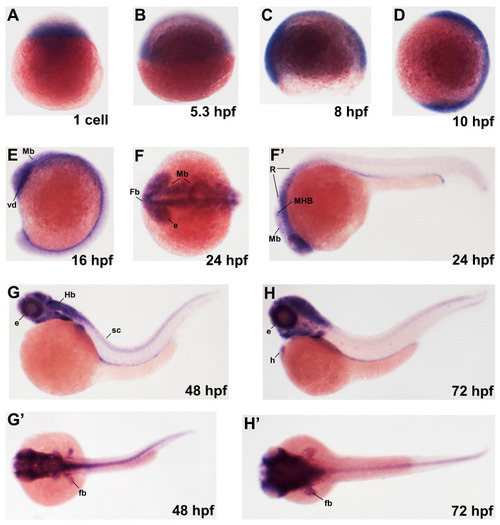
Dynamic expression of pla2g6 during zebrafish embryonic development. Whole-mount in situ hybridization analysis showing the temporal and spatial expression pattern of pla2g6 during zebrafish embryogenesis. Embryos are shown in lateral or dorsal views with anterior to the left and dorsal to the top, unless otherwise indicated. (A) Maternal pla2g6 transcripts were detected at the one-cell stage. (B–D) During blastula and early gastrula stages (5.3–10 hpf), pla2g6 expression was broadly distributed throughout the embryo. (E) At mid-segmentation stages (16 hpf), expression became enriched in the developing neural plate, including the presumptive brain and spinal cord. (F,F’) By 24 hpf, strong expression was observed in the developing brain and developing foregut and hindgut. (G,G’) At 48 hpf, pla2g6 expression was predominantly localized to brain, with detectable expression along the spinal cord. (H,H’) At 72 hpf, expression remained enriched in the brain, with additional signals detected in the developing heart and fin buds. Abbreviations: Mb, midbrain; Fb, forebrain; Hb, hindbrain; MHB, midbrain–hindbrain boundary; sc, spinal cord; e, eye; h, heart; vd, ventral diencephalon; R, rhombomere.
|
