
Fig. 3
- ID
- ZDB-FIG-260528-9
- Publication
- Ghosh et al., 2025 - EIPR1 variants cause a neurodevelopmental disorder with endolysosomal and dense core vesicle defects
- Other Figures
- All Figure Page
- Back to All Figure Page
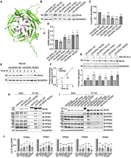
Biochemical properties of EIPR1 variant proteins. (A) Structure of human EARP-interacting protein 1 (EIPR1) (https://www.uniprot.org/) (Q53HC9) predicted using AlphaFold 2 and visualized using PyMOL version 2.5.2 (www.pymol.org). The N- and C-termini and the position of the variants R279, R16, V140, H271 and R232 residues are indicated. (B) HEK293T cells were transfected with plasmids encoding wild-type (WT) or patient variants of human EIPR1 tagged with three copies of the haemagglutinin (HA) epitope (HA-EIPR1). At 24 h post-transfection, cells were analysed by sodium dodecyl sulfate-polyacrylamide gel electrophoresis (SDS-PAGE) and immunoblotting with antibodies to the HA epitope and to glyceraldehyde-3-phosphate dehydrogenase (GAPDH) as a loading control. (C) Quantification of HA-EIPR1 protein levels normalized to GAPDH protein from three independent experiments such as that shown in B. WT HA-EIPR1 level was set to 100%. Values are the mean ± standard deviation (SD). Statistical significance was calculated by ANOVA with multiple comparisons using Dunnett’s test. ***P < 0.001. (D) HEK293T cells were transfected with plasmids encoding WT or the indicated HA-EIPR1 variants. At 24 h post-transfection, total RNA was isolated and mRNA expression measured using quantitative reverse transcription polymerase chain reaction (qRT-PCR). Graph shows the quantification of the variant mRNA expression normalized to WT expression. Values are the mean ± SD. Statistical significance was calculated by ANOVA with multiple comparisons using Dunnett’s test. *P < 0.05, ns: not significant. (E) HEK293T cells were transfected with plasmids encoding WT or R279G variant of HA-EIPR1. At 24 h post-transfection, cells were treated with 40 μM of the proteasome inhibitor MG132 for 0–6 h. Cells were subsequently analysed by SDS-PAGE and immunoblotting using antibodies to the HA epitope and to GAPDH as a loading control. (F) Quantification of HA-EIPR1 protein normalized to GAPDH protein from three independent experiments such as that shown in E. Values are the mean ± SD. Statistical significance was calculated by Student’s t-test. *P < 0.05, **P < 0.01, ns: not significant. (G) HEK293T cells were transfected with plasmids encoding WT or R279G, H271Q, R16H, V140D, R232W variants of HA-EIPR1. At 24 h post-transfection, cells were treated with 40 μM of the proteasome inhibitor MG132 for 6 h or left untreated. Cells were subsequently analysed by SDS-PAGE and immunoblotting using antibodies to the HA epitope and to GAPDH as a loading control. Quantification of HA-EIPR1 protein normalized to GAPDH protein from three independent experiments. The difference of protein accumulation was compared in individual variants between untreated and 6 h MG132 treatment. Values are the mean ± SD. Statistical significance was calculated by Student’s t-test. *P < 0.05, **P < 0.01. (H and I) HEK293T cells were transfected with plasmids encoding WT and R279G (H), or R16H, V140D, H271Q, R232W (I) variants of HA-EIPR1. At 22 h post-transfection, cell extracts were subjected to immunoprecipitation with antibody to the HA epitope conjugated to magnetic beads, followed by SDS-PAGE and immunoblotting with antibodies to the HA epitope, the indicated subunits of endosome-associated recycling protein (EARP) and GARP, and GAPDH as a loading control. (J) The interaction (IP bands) of endogenous VPS50, VPS51, VPS52, VPS53 and VPS54 with HA-EIPR1 was quantified and compared between WT and variants from three independent experiments. Values are the mean ± SD. Statistical significance was calculated by ANOVA with multiple comparisons using Dunnett’s test. *P < 0.05, **P < 0.01, ***P < 0.001. In B, E, G, H and I, the positions of molecular mass markers (in kDa) are indicated on the left. |