
Fig. 2
- ID
- ZDB-FIG-260528-8
- Publication
- Ghosh et al., 2025 - EIPR1 variants cause a neurodevelopmental disorder with endolysosomal and dense core vesicle defects
- Other Figures
- All Figure Page
- Back to All Figure Page
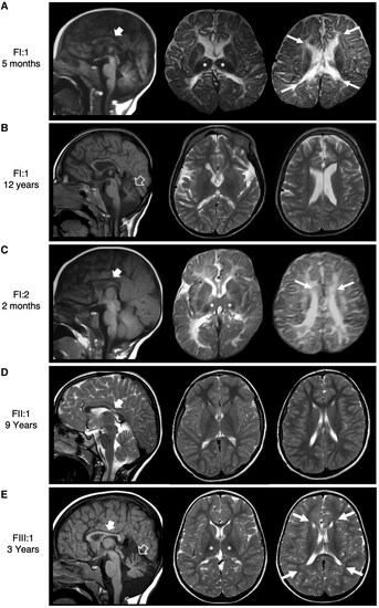
Neuroimaging findings. (A–E) Brain MRI with sagittal T1-weighted images (first column) and axial-T2-weighted images (second and third columns) performed at 6 months (A) and 12 years (B) of age for Patient FI:1, 2 months of age for Patient FI:2 (C), 9 years of age for Patient FII:1 (D) and 3 years of age for Patient FIII:1 (E). The first MRI scans in the first two subjects (Patients FI:1 and FI:2) (A and C) show a reduction of the periventricular volume with pointed appearance of the frontal and occipital horns of the lateral ventricles (arrows). There are widespread T2-signal alterations of the periventricular white matter with poor visualization of the posterior limbs of internal capsules, in keeping with delayed myelination. The corpus callosum is hypoplastic, especially in the posterior portions (thick arrows) and the anterior commissure is not visible. The thalami are small (asterisks). There is an enlargement of the cerebral subarachnoid spaces with prevalent involvement of the frontal and temporal regions. The follow-up MRI of Patient FI:1 (B) demonstrates mild cerebellar atrophy (empty arrow), while both the enlargement of the cerebral subarachnoid spaces and the volume of the periventricular white matter and thalami are stable. The signal of the white matter is normal, in keeping with completed myelination. MRI of Patient FII:1 at 9 years of age (D) shows corpus callosum hypoplasia with marked thinning of the isthmic region, and only mild white matter thinning. MRI of Patient FIII:1 at the age of 3 years (E) shows atrophy of the cerebellum with enlarged 4th ventricle and shrunken cerebellar folia and prominent fissures. |