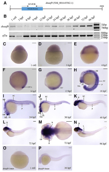
dnaaf9 exhibits a broad expression domain across the early developmental stages of zebrafish. (A) Schematic representation of dnaaf9 mRNA showing the position of primers used for WISH probe synthesis and RT-PCR analysis. (B) Temporal expression analysis of dnaaf9 mRNA during zebrafish development, using ef1α as a loading control. (C–N) Whole-mount in situ hybridisation marking dnaaf9 transcript localisation from the 1-cell to 96 hpf. (C–E) dnaaf9 is ubiquitously expressed during early development (1-cell to 6 hpf), with low maternal deposition and early zygotic expression. (F,G) Expression becomes restricted to dorsal forerunner cells (DFC) and Kupffer’s vesicle (KV) at 9 and 12 hpf, respectively. (H–J) From 18 hpf onward, dnaaf9 is detected in the telencephalon (TEL), diencephalon (DI), hindbrain (HB), pronephros (P) and spinal cord (SC). (I–N) At 30 hpf and later, expression extends to the midbrain (MB). (K–N) After 48 hpf, expression intensifies in the brain relative to other tissues. (N) By 96 hpf, expression is also detected in the liver (L). (M) Dorsal view of a 72 hpf embryo head showing dnaaf9 expression in the brain (B), retina (R), and olfactory pit (OP). (O,P) WISH using the dnaaf9 sense probe in 1 cell and 30 hpf zebrafish embryos shows no detectable signal. Scale bars, 150 µm.
|
