
Fig. 3.
- ID
- ZDB-FIG-260518-25
- Publication
- Brown et al., 2026 - Enhancer-directed gene delivery for digit regeneration based on conserved epidermal factors
- Other Figures
- All Figure Page
- Back to All Figure Page
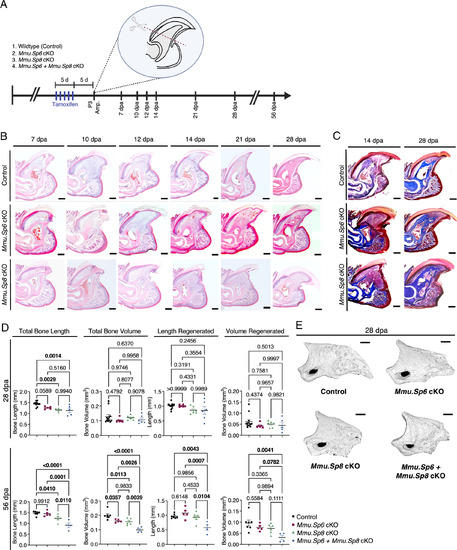
Sp6 and Sp8 are required for mouse digit tip regeneration. (A) Experimental design of Mmu.Sp6, Sp8, and Sp6+Sp8 conditional knockout models using the mouse digit tip (P3, third phalanx) amputation (amp.) model with tamoxifen induction. The timeline indicates tissue collection at 7 to 56 dpa. (B) H&E time course of digit regeneration following P3 amputation in Sp6 and Sp8 cKO mice compared with controls. Compared with controls, both cKO genotypes show delayed/defective regeneration, characterized by reduced or disorganized blastema and diminished distal bone outgrowth at indicated stages. (Scale bar, 100 μm.) (C) Masson’s trichrome staining of digit tips at 14 and 28 dpa in Sp6 cKO and Sp8 cKO mice compared with controls. (Scale bar, 100 μm.) (D) Quantification of digit tip regeneration from micro-CT analyses in tamoxifen-induced WT controls, Sp6 cKO, Sp8 cKO, and Sp6+Sp8 cKO mice at 28 and 56 dpa. Total bone length and volume, as well as regenerated length and volume, were measured. Data are presented as mean ± SEM (at 28 dpa: Control n = 11, Mmu.Sp6 cKO n = 5, Mmu.Sp8 cKO n = 5, Sp6+Sp8 cKO: n = 5; at 56 dpa: Control n = 7, Mmu.Sp6 cKO n = 5, Mmu.Sp8 cKO n = 4, Sp6+Sp8 cKO: n = 4). P-values were calculated from one-way ANOVA. (E) Representative 3D micro-CT reconstructions of digit tip regeneration in control, Sp6 cKO, Sp8 cKO, and Sp6+Sp8 cKO mice at 28 dpa. (Scale bar, 200 μm.) |