FIGURE

Figure 5.

ID
ZDB-FIG-260427-59
Publication
Gao et al., 2026 - PCYT1A Hypophosphorylation Underlies Retinal Lipid Dysregulation in CERKL Retinitis Pigmentosa and Is Therapeutically Reversed by Phosphatidylcholine
Other Figures
All Figure Page
Back to All Figure Page
Figure 5.

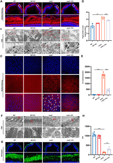
PC addition alleviates LD accumulation and photoreceptor dysfunction in cerkl−/− zebrafish. (A) Confocal imaging of LDs in WT and cerkl−/− retina with PC treatment zebrafish by Nile Red staining. Blue represents DAPI and red represents Nile Red staining. RPE, retinal pigment epithelium; ONL, outer nuclear layer; INL, inner nuclear layer. Scale bars = 10 µm. (B) TEM analysis of LDs in WT and cerkl−/− retina with PC treatment zebrafish. LD, lipid droplets. Scale bars = 10 µm. (C) Confocal imaging of LDs in WT and cerkl−/− RPE with PC treatment zebrafish by Nile Red staining. Blue represents DAPI and red represents Nile Red staining. Scale bars = 10 µm. (D) TG levels in WT and cerkl−/− retina with PC treatment zebrafish by TGs analysis kits. **, P < 0.01, ***, P < 0.001. (E) Quantitative analysis of LDs is shown in D. ***, P < 0.001. (F) TEM analysis of photoreceptors in WT and cerkl−/− retina with PC treatment zebrafish. Bars = 10 µm. (G) Immunofluorescence analysis of retinas with PC treatment zebrafish using anti-Rhodopsin antibody. Scale bars = 10 µm. (H) Quantitative analysis of the rhodopsin is shown in G. **, P < 0.01, ***, P < 0.001.

Expression Data

Expression Detail
Antibody Labeling
Phenotype Data

Phenotype Detail
Acknowledgments
This image is the copyrighted work of the attributed author or publisher, and ZFIN has permission only to display this image to its users. Additional permissions should be obtained from the applicable author or publisher of the image. Full text @ Invest. Ophthalmol. Vis. Sci.