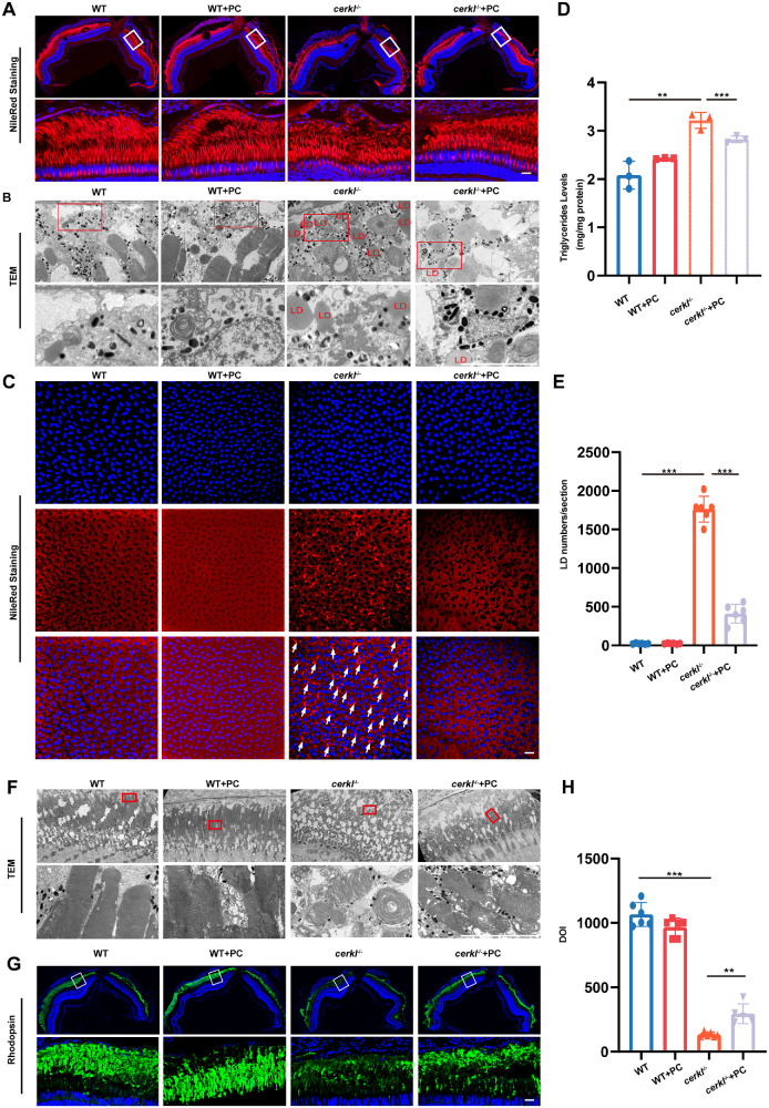
PC addition alleviates LD accumulation and photoreceptor dysfunction in cerkl−/− zebrafish. (A) Confocal imaging of LDs in WT and cerkl−/− retina with PC treatment zebrafish by Nile Red staining. Blue represents DAPI and red represents Nile Red staining. RPE, retinal pigment epithelium; ONL, outer nuclear layer; INL, inner nuclear layer. Scale bars = 10 µm. (B) TEM analysis of LDs in WT and cerkl−/− retina with PC treatment zebrafish. LD, lipid droplets. Scale bars = 10 µm. (C) Confocal imaging of LDs in WT and cerkl−/− RPE with PC treatment zebrafish by Nile Red staining. Blue represents DAPI and red represents Nile Red staining. Scale bars = 10 µm. (D) TG levels in WT and cerkl−/− retina with PC treatment zebrafish by TGs analysis kits. **, P < 0.01, ***, P < 0.001. (E) Quantitative analysis of LDs is shown in D. ***, P < 0.001. (F) TEM analysis of photoreceptors in WT and cerkl−/− retina with PC treatment zebrafish. Bars = 10 µm. (G) Immunofluorescence analysis of retinas with PC treatment zebrafish using anti-Rhodopsin antibody. Scale bars = 10 µm. (H) Quantitative analysis of the rhodopsin is shown in G. **, P < 0.01, ***, P < 0.001.
Acknowledgments
This image is the copyrighted work of the attributed author or publisher, and
ZFIN has permission only to display this image to its users.
Additional permissions should be obtained from the applicable author or publisher of the image.
Full text @ Invest. Ophthalmol. Vis. Sci.
Your Input Welcome
Thank you for submitting comments. Your input has been emailed to ZFIN curators who may contact you if
additional information is required.
Oops. Something went wrong. Please try again later.