|
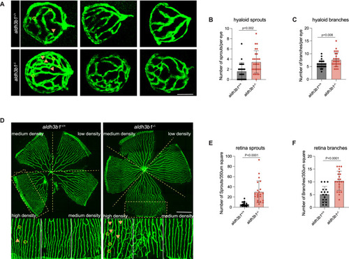
Microvascular alterations in <italic toggle='yes'>aldh3b1</italic><sup><italic toggle='yes'>-/-</italic></sup> mutants.A Representative confocal images of hyaloid vasculature revealed significant vascular alterations in aldh3b1-/- larvae at 5dpf. Yellow arrows, sprouts; yellow circles, branchpoints. White scale bar = 50 μm. B, C Quantitative analysis demonstrated a significant increase in hyaloid sprouts and branchpoints in aldh3b1-/- larvae (n = 24/27). D Representative confocal images of adult retinal vasculature showed vascular alterations in aldh3b1-/- zebrafish. Yellow arrows, sprouts; yellow circles, branchpoints. White scale bar = 500 μm. E, F Quantification of increased retinal sprouts and branchpoints in adult aldh3b1-/- zebrafish. One datapoint means one 350 μm2 square in high-density retina (n = 8/9). The bars indicate mean ± SD values. Statistical analysis was performed by a two-tailed Student’s t test. Source data are provided as a Source Data file.
|