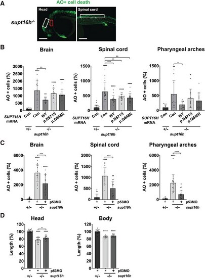
Reduced activity of SUPT16H variants in rescuing p53-mediated apoptosis in the CNS and pharyngeal arches of supt16h−/− embryos. (A) Confocal images showing acridine orange-positive apoptotic cells (green) in supt16h−/− embryos. Apoptosis was assessed at 36 hpf in the brain (white box) and pharyngeal arches (red box) of the head (left panel) and at 2 dpf in the spinal cord (white box) of the trunk (right panel). Scale bars, 200 μm. (B) Quantification of acridine orange-positive cells in the brain, pharyngeal arches, and spinal cord of supt16h+/− and supt16h−/− embryos injected with wild-type SUPT16H (WT), SUPT16H variants (c.1943A > G and c.1712A > G), or TagRFP mRNA (con). Data are presented as mean ± SD; n ≥ 7 embryos per group for head regions and n ≥ 14 for trunk regions. (C) p53 mediates apoptosis in supt16h−/− embryos. Quantification of acridine orange–positive cells in the brain, pharyngeal arches, and spinal cord at 2 dpf shows that injection of p53 morpholino (p53MO) markedly reduces apoptosis (see also Fig. S3). Data are presented as mean ± SD; n ≥ 12 embryos per group. (D) p53 knockdown alleviates craniofacial dysmorphology in supt16h−/− embryos. Data are presented as mean ± SD; n ≥ 13 embryos per group. Head size and body length were quantified from bright-field images using ImageJ in supt16h+/− and supt16h−/− embryos with or without p53MO injection. Data are presented as mean ± SD; n ≥ 13 embryos per group. Statistical significance was determined using one-way ANOVA followed by Dunnett’s multiple comparison test; *P < 0.05, **P < 0.01, ***P < 0.001, ****P < 0.0001. Abbreviations: ANOVA, analysis of variance; CNS, central nervous system; con, control (TagRFP mRNA-injected); dpf, days post-fertilization; hpf, hours post-fertilization; p53MO, p53 morpholino oligonucleotide; SD, standard deviation; SUPT16H, suppressor of ty 16 protein; supt16h, suppressor of ty 16 homolog (zebrafish gene); WT, wild type.
|
