
Fig. 1
- ID
- ZDB-FIG-260406-54
- Publication
- Lee et al., 2026 - SUPT16H-associated neurodevelopmental disorder and neurocristopathy: genetic and phenotypic spectrum
- Other Figures
- All Figure Page
- Back to All Figure Page
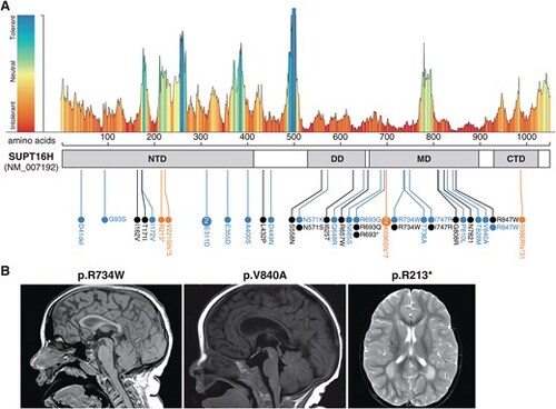
Constraint landscape of SUPT16H, variant distribution, and representative brain MRIs. (A) MetaDome tolerance plot and schematic of human SUPT16H (NM_007192), annotated with putative domains and locations of novel (blue) and previously reported (grey) missense variants, as well as novel truncating variants (orange). Domains: N-terminal domain (NTD; amino acids 5–411), dimerization domain (DD; 529–651), middle domain (MD; 660–895), and C-terminal domain (CTD; 927–1020). (B) Representative MRI scans. T1-weighted sagittal images show HCC predominantly involving the genu (individual 7, p.R734W; individual 11, p.V840A), and an axial T2-weighted image shows posterior periventricular white-matter hyperintensity (individual 13, p.R213*). Abbreviations: CTD, C-terminal domain; DD, dimerization domain; HCC, hypoplastic corpus callosum; MD, middle domain; MRI, magnetic resonance imaging; NTD, N-terminal domain; SUPT16H, suppressor of ty 16 homolog. |