FIGURE

Fig 3

ID
ZDB-FIG-260311-715
Publication
Corcoran et al., 2026 - Combined mutations of hoxa13a, hoxa13b, and hoxd13a lead to structural shifts in zebrafish soft fin rays providing insight into spiny ray evolution
Other Figures
All Figure Page
Back to All Figure Page
Fig 3

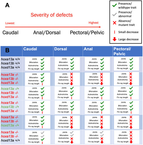
Summary of defects in each fin type for triple and double homozygous hox13 mutant fish.

(A) The caudal fin demonstrates the least severe defects in these mutants. The dorsal and anal fins lack joints, bifurcations and actinotrichia, but do not have length reductions as severe as the paired fins. The paired fins demonstrate the most severe defects in hox13 double and triple homozygous mutants. (B) A summary of observed phenotypes in double and triple homozygous mutants for the presence/ absence of joints, bifurcations and actinotrichia, as well as severity of length reductions. The genotypes of the double and triple mutants are indicated in the left column: Heterozygotes for each gene are shown in green, homozygous mutants are shown in red, and wildtypes are shown in black. Number of fish observed for each group include n = 6 for wildtype siblings, n = 8 for hoxa13a-/-, a13b-/-, d13a-/+, and n = 10 for hoxa13a-/-, a13b-/-, d13a-/-, n = 11 for hoxa13a-/-, a13b-/+, d13a-/-, n = 9 for hoxa13a-/+, a13b-/-, d13a-/-, n = 2 for hoxa13a-/-, a13b-/-, d13a+/+. Bright field image examples for each mutant are shown in Fig 4.

Expression Data

Expression Detail
Antibody Labeling
Phenotype Data
Fish:
Observed In:
Stage: Adult

Phenotype Detail
Acknowledgments
This image is the copyrighted work of the attributed author or publisher, and ZFIN has permission only to display this image to its users. Additional permissions should be obtained from the applicable author or publisher of the image. Full text @ PLoS Genet.