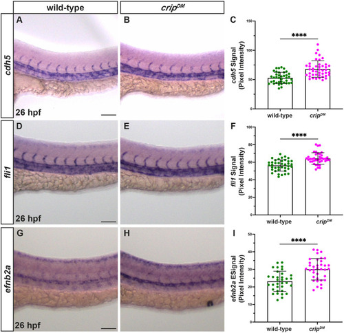
cripDM embryos exhibit upregulation of the vascular gene signature. (A-C) In situ hybridization (ISH) for cdh5 in wild-type (n=40) (A) and cripDM (n=41) (B) embryos at 26 hpf. Lateral views, anterior to the left. Quantification of cdh5 signal in the dorsal aorta (DA) using pixel intensity analysis (C) shows enhanced signal in cripDM embryos employing an unpaired nonparametric Mann–Whitney U-test (****P<0.0001). (D-F) ISH for fli1 in wild-type (n=56) (D) and cripDM (n=57) (E) embryos at 26 hpf. Lateral views, anterior to the left. Quantification of fli1 signal in the DA using pixel intensity analysis (F). An unpaired two-tailed nonparametric t-test yields a statistically significant difference between wild-type and cripDM (****P<0.0001) embryos. (G-I) ISH for efnb2a in wild-type (n=54) (G) and cripDM (n=53) (H) embryos at 26 hpf. Lateral views, anterior to the left. Quantification of efnb2a signal in the DA using pixel intensity analysis (I). An unpaired two-tailed nonparametric t-test yields a statistically significant difference between wild-type and cripDM (****P<0.0001) embryos. Mean and standard error of each dataset are shown. Scale bars: 100 µm.
|
