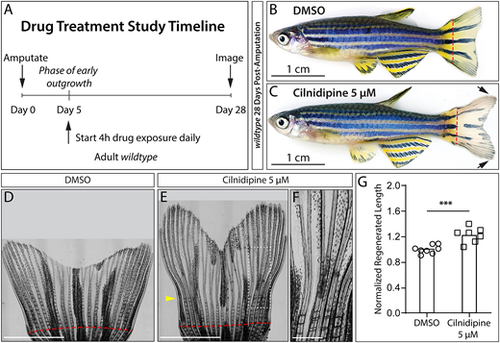
L-and/or N-type Ca2+ channels actively promote fin outgrowth cessation. (A) Timeline for outgrowth phase drug treatments of caudal fin-resected adult zebrafish from 5-28 days post amputation (dpa). (B,C) Representative bright-field images of (B) DMSO and (C) 5 µM cilnidipine-treated zebrafish at 28 dpa. The black arrows indicate overgrown regenerating caudal fin tissue. Dashed red lines mark amputation planes. Scale bars: 1 cm. (D-F) Stitched bright-field images showing 28 dpa regenerated caudal fins of the same fish in B and C. Scale bars: 4 mm. A red dashed line indicates the amputation plane. The yellow arrowhead indicates the onset of joint formation defects upon cilnidipine treatment initiation at 5 dpa. D and E are also shown in Fig. S5. (F) The zoomed region outlined in E. Scale bars: 200 µm. (G) Graph showing body length-normalized regenerated fin lengths (measured at the 3rd ray) of DMSO (control, n=8) and 5 µM cilnidipine (n=7) treated fish. Each point represents an individual fish. ***P=0.0004 by an unpaired Student's t-test.
|
