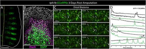
The tph1b:GCaMP6s line demonstrates Ca2+ fluxes in distal blastemal cells of regenerating fins. (A) Whole-mount confocal image of a 3 day post amputation (dpa) regenerating caudal fin of a tph1b:GCaMP6s (green) adult fish. The yellow dashed line shows the amputation plane. The fin is outlined. Scale bar: 500 µm. (B) tph1b-driven GCaMP6s expression includes distal blastemal cells. Confocal maximum intensity projection image of an antibody-stained 3 dpa tph1b:GCaMP6s regenerating fin section (representative of n=5 fins). Dachshund (Dach) and EGFP (for GCaMP6s) are in magenta and green, respectively. Hoechst-stained nuclei are gray. Scale bar: 50 μm. (C) Frames from a confocal movie showing the distal blastema of a tph1b:GCaMP6s regenerating caudal fin at 3 dpa (representative of more than 20 fish observed over six imaging sessions). Scale bars: 50 µm. The arrows indicate selected cells with dynamic GCaMP6s fluorescence over the 7-min period. The large arrows indicate the same cells during GCaMP6s-reported Ca2+ spikes. (D) Plots of normalized GCaMP6s intensity over time for the numbered cells in C (green traces) and additional cells (black traces) showing heterogenous Ca2+ dynamics over the 7-min period.
|
