
FIGURE 7
- ID
- ZDB-FIG-251115-169
- Publication
- Bolanos-Palmieri et al., 2025 - Kynurenine Pathway Dysregulation Impairs Podocyte Morphology and Bioenergetics In Vitro and Leads to Glomerular Dysfunction
- Other Figures
- All Figure Page
- Back to All Figure Page
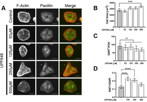
KMO inhibition impacts cell shape, size and proliferation in cultured murine parietal epithelial cells. Mouse parietal epithelial cells (PEC) were seeded on glass cover slides and treated with the KMO inhibitor UPF648 or ethanol control for 48 h at the concentrations stated in the figure. (A) Representative images of mouse PECs after KMO inhibition. The focal adhesions (paxillin in green) and Actin filaments (phalloidin in red) are shown. Scale bar 50 μm. Photos were taken at 40X. (B) Quantification of changes in cell size after KMO inhibition. Total cell area in μm2 was quantified from over 80 photographs taken at 40X of phalloidin‐labeled PECs. Bars represent mean cell area ±95% CI. Data from two independent experiments (C) Average number of DAPI+ nuclei per field of view (FOV). The cells were photographed at 10X and all nuclei were counted. Twenty photographs were taken for each group, data from two independent experiments. Bars represent the mean cell number per FOV ±95% CI. (** |