
FIGURE 3
- ID
- ZDB-FIG-251115-165
- Publication
- Bolanos-Palmieri et al., 2025 - Kynurenine Pathway Dysregulation Impairs Podocyte Morphology and Bioenergetics In Vitro and Leads to Glomerular Dysfunction
- Other Figures
- All Figure Page
- Back to All Figure Page
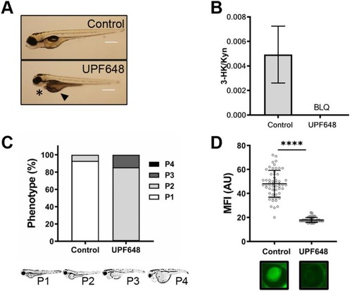
Inhibition of Kmo by UPF648 also leads to the development of hallmarks of proteinuric kidney disease in zebrafish larvae. Zebrafish larvae were treated with the Kmo inhibitor UPF648 at a concentration of 100 μM or with EtOH control after the hatching period (48 hpf) and for the following 48 h. Exposure to the chemicals was done through immersion treatment. Larval development was monitored until 96 hpf and readouts of edema formation and proteinuria were collected. (A) Representative image of the zebrafish larvae after inhibition of Kmo activity by treatment with UPF648. A higher proportion of larvae show signs of pericardial effusion (*) and yolk sac edema (▲) when compared to controls. (B) A reduction in the 3‐HK/KYN ratio indicates a reduced catabolism of kynurenine by Kmo after inhibition with UPF648. All metabolite quantifications have been corrected for variations in protein content in each group, data collected from two independent experiments, each with 10 fish per treatment. BLQ = below level of quantification. (C) Quantification of the proportion of larvae belonging to each of the phenotypic categories based on the severity of the generalized edema. (D) A reduction in maximum fluorescence intensity (MFI) measurement shows that the larvae develop proteinuria upon Kmo inhibition by UPF648. Below the graph denoting the MFI quantification is a representative photograph of the retinal vessel plexus of a larva for each group ( |
| Fish: | |
|---|---|
| Condition: | |
| Observed In: | |
| Stage: | Day 4 |