
Fig. 6
- ID
- ZDB-FIG-251107-62
- Publication
- Leach et al., 2025 - Macrophage/microglia-dependent mechanisms drive retinal pigment epithelium regeneration in zebrafish
- Other Figures
- All Figure Page
- Back to All Figure Page
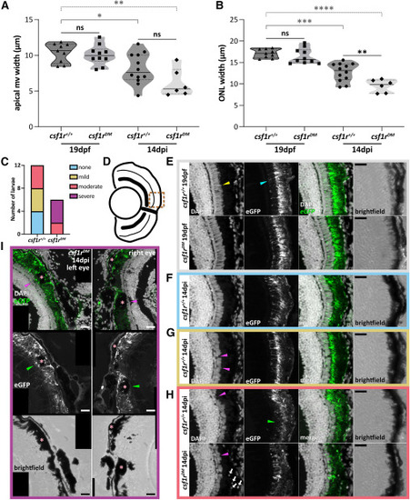
csf1rDM zebrafish fail to regenerate by 14 dpi (A and B) Violin plots showing (A) RPE apical microvilli (mv) and (B) photoreceptor (ONL) width measurements in 19 dpf unablated (MTZ–) and 14 dpi ablated (MTZ+) larvae. (C) Stacked column graph showing the degree of phenotype severity in 14 dpi larvae (n = 12 total, csf1r+/+; n = 6 total, csf1rDM). (D) Schematic of a late-stage larval eye depicting the central-dorsal region zoomed in on in (E–I) (bronze dotted line). (E) Transverse confocal micrograph digital zooms showing 19 dpf unablated (MTZ–) csf1r+/+ and csf1rDM larvae. Examples of uniformly normal photoreceptor (elongated outer segments) and RPE (apical microvilli) morphologies are highlighted with yellow and cyan arrowheads, respectively. (F–H) Transverse confocal micrograph digital zooms showing 14 dpi ablated (MTZ+) csf1r+/+ or csf1rDM larvae with (F) no discernible phenotype, (G) mild ONL and RPE phenotypes, and (H) moderate ONL and RPE phenotypes. Magenta arrowheads point out areas of sparse, absent, or hypertrophic outer segments. Green arrowhead indicates an area where apical microvilli are missing or truncated. White arrows indicate cellular debris retention. (I) Stitched transverse confocal micrographs showing the right and left eyes of a 14 dpi ablated (MTZ+) csf1rDM larva with severe ONL and RPE phenotypes. Green and magenta arrowheads point to areas of abnormal RPE and photoreceptor outer segments, respectively, that localize with large, pigmented debris deposits (asterisks). DAPI = nuclei (white); rpe65a:nfsB-eGFP = RPE (white and green). Scale bars, 20 μm; ns, not significant; ∗p ≤ 0.05; ∗∗p ≤ 0.01; ∗∗∗p ≤ 0.001; and ∗∗∗∗p ≤ 0.0001. Experimental replicates and statistical information can be found in Table S3. |