
Fig. 4
- ID
- ZDB-FIG-251107-60
- Publication
- Leach et al., 2025 - Macrophage/microglia-dependent mechanisms drive retinal pigment epithelium regeneration in zebrafish
- Other Figures
- All Figure Page
- Back to All Figure Page
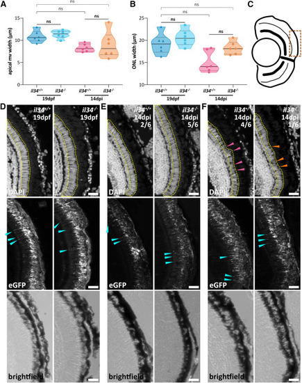
il34 mutants regenerate by 14 dpi despite early time point deficiencies (A and B) Violin plots showing (A) RPE apical microvilli (mv) and (B) photoreceptor (ONL) width measurements in 19 dpf unablated (MTZ–) and 14 dpi ablated (MTZ+) larvae. (C) Schematic of a late-stage larval eye depicting the central-dorsal region zoomed in on in (D–F) (bronze dotted line). (D and E) Transverse confocal micrograph digital zooms showing (D) 19 dpf unablated (MTZ–) il34+/+ and il34−/− larvae and (E) 14 dpi ablated (MTZ+) il34+/+ and il34−/− animals with no discernible phenotype. Examples of uniformly normal photoreceptors (elongated outer segments) and RPE (apical microvilli) morphologies are highlighted with yellow dotted lines and cyan arrowheads, respectively. (F) Digital zooms showing 14 dpi ablated (MTZ+) il34+/+ and il34−/− larvae with normal RPE but truncated or disorganized outer segments (arrowheads point to examples). All scale bars, 20 μm; DAPI = nuclei (white); rpe65a:nfsB-eGFP = RPE (white and green); ns, not significant. Experimental replicate and statistical information can be found in Table S3. |