
Fig. 1
- ID
- ZDB-FIG-251023-1
- Publication
- Méndez-Martínez et al., 2025 - Genetic ablation of Pth4 disrupts calcium-phosphate balance, bone development, and kidney transcriptome in teleosts
- Other Figures
- All Figure Page
- Back to All Figure Page
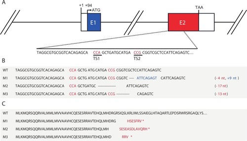
CRISPR/Cas9-induced mutations at the zebrafish pth4 locus. (A) Graphical representation of the pth4 gene. Coding exons are represented with colored boxes and the 5′-UTR and 3′UTR are represented as white boxes. (B) Nucleotide sequence of wild-type pth4 (WT) and induced mutations (M1, M2 and M3). The protospacer-adjacent motif (PAM) is indicated in red, denoting the mutation target sites (TS1 and TS2). Hyphens represent deletions and nucleotides in blue represent insertions. (C) Predicted amino acid sequences of wild-type pth4 and induced mutations. Amino acids in red differ from the WT sequence and asterisks indicate stop codons. (For interpretation of the references to color in this figure legend, the reader is referred to the web version of this article.) |