
Fig. 13
- ID
- ZDB-FIG-251015-19
- Publication
- Ota et al., 2025 - Evolutionary Insights into Muscle Fiber Distribution in the Twin Tails of Ornamental Goldfish
- Other Figures
- All Figure Page
- Back to All Figure Page
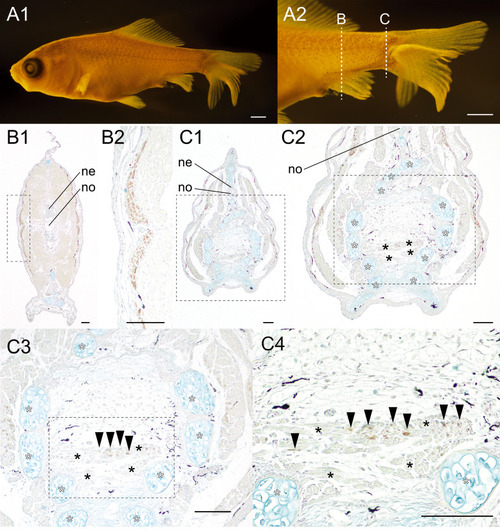
Distribution pattern of the slow muscle fibers at the 24 twin #01 of the lab goldfish strain. (A). The whole body (A1) and the magnified view of the caudal region (A2) of the goldfish (#2024-0419-03-#01, 13.07 mm [SL], 23 dpf). (B–C). Different levels of the sections immunostained with the slow muscle fiber specific antibody (F59). The magnified views of the sections are indicated by dashed line boxes with panel labels. White asterisks indicate the ventral caudal skeleton complex including pural, hypural, and hemal spines. The medial caudal muscle fibers are indicated by black asterisks in panels C2, C3, and C4. The black arrowhead in panel D3 indicates slow muscle fibers in the medial caudal muscle. Abbreviations: ne, neural tube, no, notochord. Scale bars: A1 = 1 mm; A2, B, C = 100 μm. |