
Fig. 4
- ID
- ZDB-FIG-251015-10
- Publication
- Ota et al., 2025 - Evolutionary Insights into Muscle Fiber Distribution in the Twin Tails of Ornamental Goldfish
- Other Figures
- All Figure Page
- Back to All Figure Page
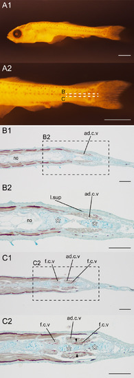
Horizontal section of the caudal region of a wild-type goldfish larva at pelvic fin bud stage. (A). The whole (A1) and the magnified (A2) view of the gold- fish (#2020-0406-01C, 8.07 mm [SL], 26 dpf). (B, C). Sections immunostained with the slow muscle fiber specific antibody (F59) in the caudal region. The sectioned levels are indicated in panel A2. The magnified views of B1 and C1 are shown in B2 and C2, respectively. White asterisks in B2 and C2 indicate axial skeleton. Slow muscle fibers of flexor caudals ventralis are indicated by black arrowheads. Abbreviations: ad.c.v, Adductor caudalis ventralis; f.c.v, flexor caudalis ventralis; l.sup, lateralis superficials. Scale bars: A = 1 mm; B, C = 100 μm. |