
Fig. 8
- ID
- ZDB-FIG-251015-14
- Publication
- Ota et al., 2025 - Evolutionary Insights into Muscle Fiber Distribution in the Twin Tails of Ornamental Goldfish
- Other Figures
- All Figure Page
- Back to All Figure Page
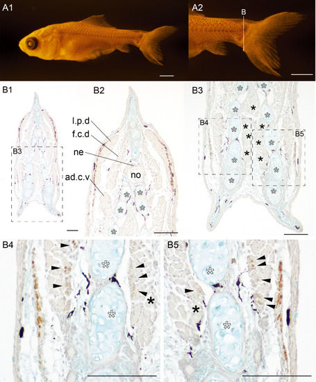
Distribution pattern of the slow muscle fibers in the Oranda strain gold-fish. (A). Whole body (A1), and magnified lateral (A2) of goldfish (#2024-0319-01-#03, 7.93 mm [SL], 31 dpf). The section at the caudal region was immunostained by the slow muscle fiber specific antibody (F59). (B). The sectioned levels are indicated in A2. White asterisks indicate the ventral caudal skeleton complex including pural, hypural, and hermal spines. Black asterisks indicate the medial caudal muscle. Abbreviations: ad.c.v, adductor caudalis ventralis; f.c.d, flexor caudalis dorsalis; l.p.d, lateralis profundus dorsalis; no, notochord; ne, neural tube. Scale bars: A1, A2 = 1 mm; B = 100 μm. |