
Fig. 6
- ID
- ZDB-FIG-251015-12
- Publication
- Ota et al., 2025 - Evolutionary Insights into Muscle Fiber Distribution in the Twin Tails of Ornamental Goldfish
- Other Figures
- All Figure Page
- Back to All Figure Page
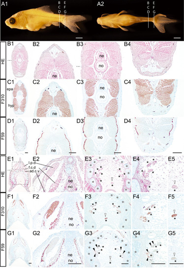
Transverse view of the trunk region of a twin-tail goldfish larva at the pelvic fin ray stage.(A). The lateral view (A1) and ventral view (A2) of Ryukin goldfish sample (#2022-0502-21-26dpdf-RY@02-0802-1A-6C-2A-#01, 8.84 mm [SL], 26 dpf). (B–G). Transverse sections hematoxylineosin stained and immunostained with the fast muscle fiber specific antibody (F310), and the slow muscle fiber specific antibody (F59) at post-anal fin level (B–D) and caudal fin level (E-G). Panels in the second to fourth columns at post-anal fin levels show magnified views of the dorsal (B2, C2, D2), mid (B3, C2, D3), and ventral (B4, C4, D4) regions. Panels in the second and third columns at caudal levels showed magnified views of the dorsal (E2, F2, G2), and ventral (E3, F3, G3) regions. Magnified areas are outlined by dashed boxes in panel E1. Panels in the fourth and fifth columns at caudal levels showed high-magnified views of the left-ventral (E4, F4, G4) and mid-ventral (E5, F5, G5) regions. Horizontal myoseptum is identified by dashed lines in panels B1, B3 and E1. The muscular tissues located between the bifurcated caudal fin skeleton are indicated by black asterisks. White asterisks indicate the ventral caudal skeleton complex including pural, hypural, and hermal spines. Black arrowheads indicate slow muscle fibers in the flexor caudalis ventralis. Abbreviations: ad.c.v, adductor caudalis ventralis; epa, epaxial muscle; f.c.d, flexor caudalis dorsalis; f.c.v, flexor caudalis ventralis; hyp, hypaxial muscle; l.p.d, lateralis profundus dorsalis; ne, neural tube; no, notochord. Scale bars: A2 = 1 mm; D1, D2, D3, D4, G1 G2, G3, G4, G5 = 100 μm. Adjacent histological sections in the same column have the same magnification. |