|
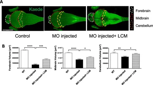
LCM restores brain size in AGTPBP1 MO-injected zebrafish embryos. A Confocal imaging of embryonic zebrafish brain at 72 hpf. Kaede was shown in green. White dotted line: forebrain; red: midbrain; and yellow: cerebellum. B Quantification of brain volume, including the forebrain, midbrain, and cerebellum before and after LCM treatment. Forebrain size of WT and MO-injected embryos (n = 3, p < 0.0001); forebrain size of MO-injected embryos with and without LCM treatment (n = 3, p = 0.0003); midbrain size of WT and MO-injected embryos (n = 3, p < 0.0001); midbrain size of MO-injected embryos with and without LCM treatment (n = 3, p = 0.0442); cerebellum size of WT and MO-injected embryos (n = 3, p = 0.0061); cerebellum size of MO-injected embryos with and without LCM treatment (n = 3, p = 0.0214). *p < 0.05, **p < 0.01, ***p < 0.001, ****p < 0.0001. Data were analyzed by one-way ANOVA, followed Tukey’s test. Data were presented as mean ± SEM
|