FIGURE

Figure 4

ID
ZDB-FIG-250816-57
Publication
Tata et al., 2025 - Prostaglandin Analogs and Eupatilin as Treatments for Nephronophthisis
Other Figures
All Figure Page
Back to All Figure Page
Figure 4

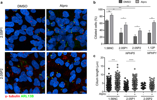
Ciliogenesis defects in NPHP5 URECs are rescued by Alprostadil/PGE1. (a) Immunofluorescence for primary cilia (ARL13B, green) and basal bodies markers (γ- tubulin, red) of NPHP5 URECs (2.05P1, 2.05P2) treated with 0.04% DMSO or 2 μM alprostadil (Alpro) for 48 hours. Scale bar 10 μm. (b) Ciliogenesis was quantified in control (1.56NC), NPHP5 (2.05P1, 2.05P2) and NPHP1 (1.12P) URECs treated with either DMSO (0.04%) or 2 μM alprostadil (Alpro), were fixed and stained similarly as in (a) (n = 4 experiments). Mixed linear regression with quasi-binomial penalization: ∗∗∗P < 0.001, ∗∗P < 0.01, ∗P < 0.05. (c) Cilium length was quantified as in Figure 1 in control (1.56NC) and NPHP5 (2.05P1, 2.05P2) URECs treated with 0.04% DMSO (-) or 2μM alprostadil (Alpro). n = 4 experiments; paired 2-tailed t: ∗∗∗∗P < 0.0001, ∗∗∗P < 0.001, ∗∗P < 0.01. DMSO, dimethyl sulfoxide; UREC, urinary renal epithelial cells.

Expression Data

Expression Detail
Antibody Labeling
Phenotype Data

Phenotype Detail
Acknowledgments
This image is the copyrighted work of the attributed author or publisher, and ZFIN has permission only to display this image to its users. Additional permissions should be obtained from the applicable author or publisher of the image. Full text @ Kidney Int Rep