
Fig. 1
- ID
- ZDB-FIG-250804-31
- Publication
- Tian et al., 2025 - Binocular integration of prey stimuli in the zebrafish visual system
- Other Figures
- All Figure Page
- Back to All Figure Page
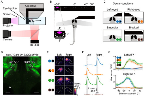
Reversible occlusion of one eye blocks the response to prey stimuli (A) Schematic of the experimental setup: the eye-blocker is positioned in front of the fish to block visual input. (B) Bright UV circulating prey-like stimuli were presented at azimuths from −50° (left) to +50° (right). (C) Larvae were tested under 4 ocular conditions: right eye blocked, left eye blocked, neither eye blocked, and both eyes blocked. (D) Baseline fluorescence image showing AF7s labeled with GCaMP6s (scale bar, 50 μm). (E) Activation of AF7 in left eye, right eye, binocular, and fully blocked conditions in one example larva (scale bar, 50 μm). Pixels are color-coded by trial-averaged increase in mean ΔF/F during the stimulus interval, compared with that during the 2 s before the stimulus. (F) Response of the left and right AF7 to 0° prey stimulus. Vertical dashed lines indicate the stimulus interval. Solid line and shaded region represent the mean and 95% confidence interval, respectively (n = 7 fish). (G) Spatial tuning curves showing maximum activity of AF7 (within the 3-s time window after stimulus onset) in response to prey stimuli at different azimuths. Solid lines and shaded regions represent the mean and 95% confidence interval, respectively (n = 7 fish). See also Figure S1. |