|
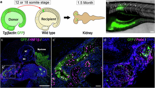
Transplanted GFP+ somite contributed to mesonephric nephrons in recipient fish. a Schematic of somite transplantation showing a GFP+ donor somite is harvested at 12 (15 hpf) or 18 (18 hpf) somite stage and transplanted into a wild-type recipient embryo at equivalent developmental stage, and kidney of the recipient fish is examined at 1.5-month of the age. b Image of a recipient larvae (6 dpf) developed donor derived GFP+ muscle fibers at the transplantation site (somite 3, transplanted at 12 somite stage). Scale bar, 20 µm. c Cross section of same recipient juvenile fish (at 1.5-month stage) immunostained for GFP and renal tubular marker Hnf1b, showing donor somite contributed tubular structures (Scale bar, 100 µm). c’ Close-up image of the area boxed in c showing Hnf1b+ mesonephric tubules containing donor somite derived GFP+ cells (Scale bar, 20 µm). Similar results were observed in 3 biologically independent recipients with positive contribution (n = 3). d Immunostaining of donor derived Podxl+/GFP+ glomeruli, Scale bar, 20 µm. Similar results were observed in 3 biologically independent recipients with positive contribution (n = 3). For merged images, levels adjustments were applied independently to individual colour channels for clarity. All adjustments were applied equally across the entire image.
|