FIGURE

Figure 5

ID
ZDB-FIG-250729-23
Publication
Shen et al., 2025 - Regulation of Hindbrain Vascular Development by rps20 in Zebrafish
Other Figures
All Figure Page
Back to All Figure Page
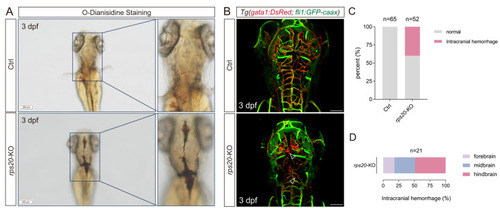
Figure 5

Knockout of rps20 caused intracranial hemorrhage. (A) Representative images of O-dianisidine staining of rps20 knockout and control zebrafish; (B) Representative images of intracranial hemorrhage of rps20 knockout zebrafish with dual-labeled vasculature and erythrocytes. Empty arrowheads indicate sites of erythrocyte aggregation outside the cerebral blood vessels; (C) Intracranial hemorrhage incidence statistics in control versus rps20-KO zebrafish. 65 WT larvae and 52 rps20-KO larvae were analyzed; (D) Regional hemorrhage distribution analysis (forebrain/midbrain/hindbrain) in rps20-KO zebrafish. A total of 21 rps20-KO larvae with intracranial hemorrhage were analyzed. Scale bar, 200 μm (A). Scale bar, 100 μm (B).

Expression Data

Expression Detail
Antibody Labeling
Phenotype Data

Phenotype Detail
Acknowledgments
This image is the copyrighted work of the attributed author or publisher, and ZFIN has permission only to display this image to its users. Additional permissions should be obtained from the applicable author or publisher of the image. Full text @ Cells