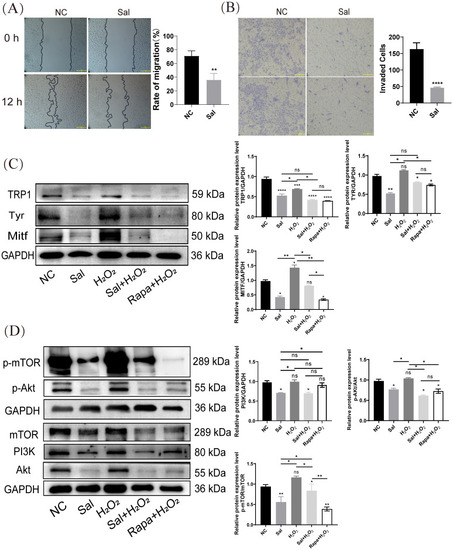
Salidroside inhibits H2O2-inductd melanogenesis by inactivating PI3K/AKT/mTOR. (A) Wounding-healing assays conducted in B16F10 cells. Cells were treated with salidroside (Sal) in serum-free 1640 medium. The wounds were photographed at 0 and 12 h after salidroside treatment. Representative photos from each group are presented in the left panels, migration rates are shown in the right panels. Data are shown as mean ± SD of three independent experiments, **p < 0.01, n = 3, scale bar = 100 μm. (B) After 24 h of salidroside treatment, B16F10 cells was detected by Trans-well assay. ****p < 0.001, n = 3, scale bar = 100 μm. (C) The protein-expression levels of melanin synthesis-related proteins (TRP1, Tyr and Mitf) in B16F10 cells were detected by western blotting. (D) The protein-expression levels of PI3K/Akt/mTOR signaling pathway-related proteins (PI3K, Akt, and mTOR) in B16F10 cells were detected by western blotting. GAPDH functioned as a loading control. NC: untreated control; Sal: salidroside; H2O2 + salidroside: After H2O2 induction for 4 h, incubation with salidroside (100 μM) was performed for 24 h; H2O2 + Rapa: After H2O2 induction for 4 h, incubation with rapamycin (2 μM) was performed for 24 (h) ns, no significance, *p < 0.05, **p < 0.01, vs. control group. Results are denoted as mean ± SD. GAPDH, glyceraldehyde 3-phosphate dehydrogenase; SD, standard deviation.
|
