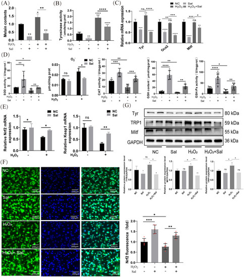
Salidroside inhibits H2O2-induced melanogenesis by promoting antioxidant effect in B16F10 cells. (A) Salidroside treatment for 24 h reduced melanin synthesis after H2O2 induction. *p < 0.05, **p < 0.01, n=3 (B) Cellular tyrosinase activity and tyrosinase activity levels were measured by dopachrome formation from L-DOPA as a substrate. (C) The mRNA expression levels of Tyr, Mitf and Oca2 in B16F10 cells were measured by RT-qPCR. ns, no significance, *p < 0.05, **p < 0.01, *** p < 0.005, ****p < 0.001, n=3. (D) B16F10 cells were treated with salidroside for 24 h or H2O2 for 4 h and cell homogenates were obtained using an ultrasonic crusher. The levels of SOD, O2•− content, CAT, GSH and GSH-px in the cells were measured by the corresponding kits. All data are expressed as mean ± SD (n = 6/9). ns, no significance, *p < 0.05, **p < 0.01, ***p < 0.005, ****p < 0.001. (E) The mRNA expression levels of Nrf2 and Keap1 in B16F10 cells were determined by RT-qPCR. ns, no significance, *p < 0.05, **p < 0.01. (F) Immunofluorescence staining results of Nrf2 in B16F10 cells (n=4), scale bar=200 μm. **p < 0.01, ***p < 0.005. (G) The protein-expression levels of Tyr, TRP-1, and Mitf were examined by western blotting analysis and greyscale analysis of proteins. GAPDH functioned as a loading control. NC, untreated control; Sal, salidroside; H2O2 + salidroside, After H2O2 induction for 4 h ns, no significance, *p < 0.05, **p < 0.01. Results were denoted as mean ± SD of three times. L-DOPA, l-3,4-dihydroxyphenylalanine; SOD, superoxide dismutase; O2•−, superoxide anion; CAT, catalase; GSH, glutathione; GSH-Px, Glutathione peroxidase; RT-qPCR, quantitative reverse transcription polymerase chain reaction; GAPDH, glyceraldehyde 3-phosphate dehydrogenase; SD, standard deviation.
|
