
Fig. 2
- ID
- ZDB-FIG-250722-53
- Publication
- Fehilly et al., 2025 - Germline Disruption of Retinal Pigment Epithelium-Expressed Zebrafish rlbp1b-/- Results in Selective Dim Light Visual Behavior Deficits and Provides a Screening Platform for Evaluating the Pathogenicity of Human RLBP1 Variants
- Other Figures
- All Figure Page
- Back to All Figure Page
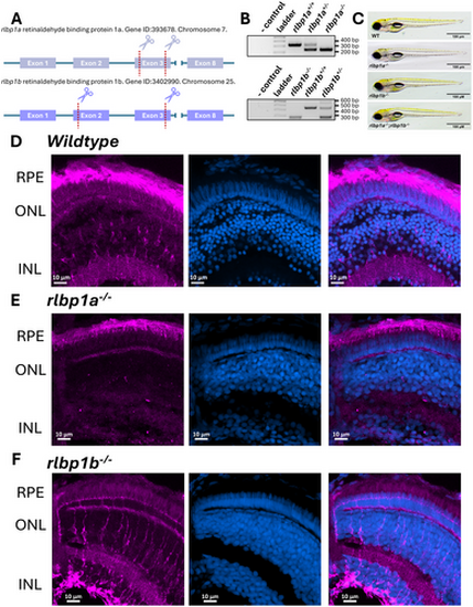
Generation and confirmation of zebrafish Cralbp CRISPR knockout lines. (A) Schematic of CRISPR approach used to generate rlbp1a and rlbp1b knockout lines. (B) Representative genotyping gels for PCR genotyping of rlbp1a and rlbp1b knockout lines where amplicons of 346 bp and 245 bp (rlbp1a) or 469 bp and 248 bp (rlbp1b) represent wildtype and deletion alleles, respectively. (C) Brightfield images of rlbp1a, or rlbp1b, and double knockout lines. (D) Cralbp immunostaining in 5 dpf larval zebrafish eyes showing expression in Müller glia and RPE. (E) Cralbp immunostaining in the rlbp1a knockout line showing loss of expression in Müller glia and robust expression in the RPE. (F) Cralbp immunostaining shows the opposite pattern in the rlbp1b knockout line with loss of expression in the RPE but persistent expression in the Müller glia. Magenta staining represents staining with the RLBP1 antibody (15356-1-AP), blue represents staining with Hoechst for nuclei. |