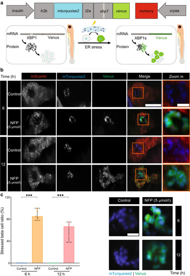
Exocrine damage induces ER stress in beta cells. (a) Schematic showing the ER stress reporter under the beta cell-specific insulin promoter; venus is fused with xbp1, which under normal conditions remains unspliced, resulting in only mTurquoise2+ nuclei. Upon ER stress, XBP1s is produced with Venus being in frame and causing stressed beta cells to display mTurquoise2+ nuclei and Venus+ cytosol in Tg(insulin:h2b-mturquoise.2-xbp1-venus;cryaa:mcherry) larvae, briefly Tg(insulin:xbp1v). Additionally, mcherry under the cryaa promoter is included to facilitate the selection of positive transient zebrafish. (b) The expression of mTurquoise2 and Venus in beta cells after exocrine damage stimulation by 5 µmol/l NFP. Scale bar, 50 μm or 10 µm (magnified image). (c) Quantification of the ratio of stress beta cells to the total number of beta cells after 6 and 12 h of NFP treatment (n=5 each). The representative images were used for quantification. Scale bar, 10 µm. Data represent the median values with bars indicating the range from the maximum to the minimum data points. Unpaired t test was used for statistical differences between groups. ***p<0.001. Part of Fig. 3a is created in BioRender. Faraj, N. (2025) https://BioRender.com/z06r241
|
