
Fig. 4
- ID
- ZDB-FIG-250714-13
- Publication
- Ning et al., 2025 - BCAS2 promotes primitive hematopoiesis by sequestering β-catenin within the nucleus
- Other Figures
-
- Fig. 1
- Fig. 1 - Supplemental 1
- Fig. 1 - Supplemental 2
- Fig. 1 - Supplemental 3
- Fig. 1 - Supplemental 4
- Fig. 2
- Fig. 2 - Supplemental 1
- Fig. 3
- Fig. 3 - Supplemental 1
- Fig. 3 - Supplemental 2
- Fig. 3 - Supplemental 3
- Fig. 3 - Supplemental 4
- Fig. 4
- Fig. 5
- Fig. 5 - Supplemental 1
- Fig. 5 - Supplemental 2
- Fig. 6
- Fig. 6 - Supplemental 1
- Fig. 7
- Fig. 7 - Supplemental 1
- Fig. 7 - Supplemental 2
- All Figure Page
- Back to All Figure Page
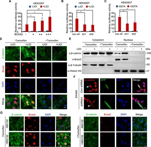
BCAS2 is essential for β-catenin nuclear accumulation. (A–C) BCAS2 enhances LiCl-induced TOPflash activity in HEK293T cells. Cells were transfected with BCAS2 expression plasmids (A), shRNA plasmids (B), or S37A-β-catenin expression plasmids (C), together with the TOPflash luciferase and Renilla luciferase vectors. After transfection, cells were subsequently treated with or without 100 ng/ml LiCl for 12 h and assayed for luciferase activity (n=3). *p<0.05; **p<0.01 (Student’s t-test). (D, E) Bcas2-cKO mouse embryonic fibroblasts (MEFs) were incubated with tamoxifen for 24 h and then treated with or without 100 ng/mL LiCl. The nuclear accumulation of β-catenin was analyzed using immunofluorescence (D) and western blotting (E). (F) SW480 cells were transfected with the indicated shRNA constructs, and the endogenous β-catenin protein was detected using immunofluorescence 48 h after transfection. The expression of GFP served as a transfection control. The arrowheads indicate the cells transfected with indicated shRNA constructs. (G) Bcas2-cKO MEFs were cultured in the presence of tamoxifen for 24 h and then treated with 20 μM MG132 for 6 h. The expression of BCAS2 and β-catenin was measured by immunofluorescence. Scale bars, 10 μm (D, F, G). |