
Fig. 5
- ID
- ZDB-FIG-250528-23
- Publication
- Kaus-Drobek et al., 2025 - From discovery to potential application: engineering a novel M23 peptidase to combat Listeria monocytogenes
- Other Figures
- All Figure Page
- Back to All Figure Page
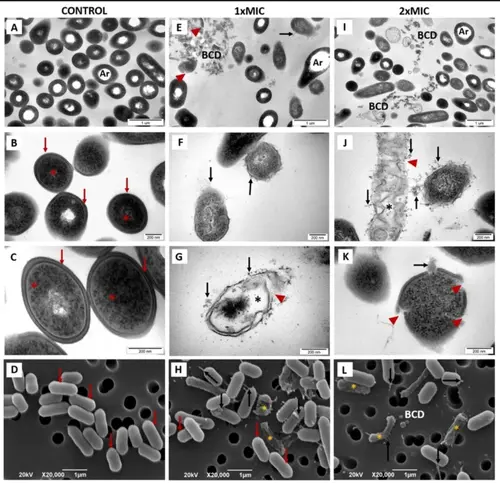
Lytic activity of the StM23_CWT protein on L. monocytogenes MF7703, as demonstrated by transmission (TEM, upper rows) and scanning (SEM, bottom row) electron microscopy studies. In the control group, no ultrastructural changes were observed, with electron micrographs showing intact, complete bacterial cell walls (A-D, red arrows) and electron-dense cytoplasmic content (B, C, red stars). In contrast, cells treated with StM23_CWT (E-L) exhibited a wide range of ultrastructural alterations. At both 1xMIC (E-H) and 2xMIC (I-L), significant bacterial cell damage was noted, including cell wall disruption (E, G, J, K, red arrowheads), leakage of cytoplasm (E- L, black arrows), accumulation of bacterial cell debris - BCD (E, I), loss of cytoplasm resulting in presence of cytoplasmic clear zones (G, J, black stars). In SEM analysis, at 1xMIC (H) group, only a few well-preserved cells were noted (red arrows), while the majority exhibited severe structural damage, including shrinkage (yellow stars) and cytoplasmic leakage (black arrows). At 2xMIC concentration (L), all cells displayed ultrastructural alterations (shrinkage and cytoplasmic leakage) and bacterial cell debris accumulation (L). Some artifacts- Ar (A, E, I), caused by sectioning procedure were comparable in all groups. |