
Fig. 9
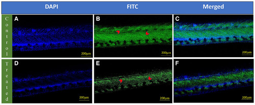
Whole-mount localization of vascular endothelial growth factor A (VEGFA) in the intersegmental vessels of 72 hpf zebrafish larvae. The upper panel (A-C) shows images of control zebrafish larvae: (A) Nuclei stained with DAPI (blue), (B) VEGFA immunolabeling visualized using FITC (green), with red arrowheads indicating VEGFA expression in the intersegmental blood vessels and (C) merged image combining DAPI and VEGFA signals. The lower panel (D–F) shows images of etoricoxib-treated zebrafish larvae: (D) Nuclei stained with DAPI (blue), (E) VEGFA immunolabeling visualized using FITC (green), with red arrowheads indicating VEGFA expression in the intersegmental blood vessels and (F) merged image combining DAPI and VEGFA signals. |