|
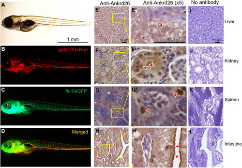
Morphology of zebrafish larvae and immunohistochemical detection of Ankrd26 protein in adult zebrafish tissues. (A-D) Representative morphology of a 4 dpf larva, including a light microscopic image, a red fluorescent (gata-1/DsRed) image, a green fluorescent (fli-1/eGFP) image and a merged image as indicated. (E-H) Immunohistochemical staining of Ankrd26 protein in hepatocytes in the liver (E,E′), proximal or distal tubules in the kidney (F,F′), ellipsoids in the spleen (G,G′) and epithelial cells in the intestinal villi (H,H′). Red arrowheads indicate representative Ankrd26-postivie cells. (I-L) Negative controls for immunohistochemical staining in the liver (I), kidney (J), spleen (K) and intestine (L), without incubation with primary antibody against zebrafish Ankrd26.
|