
Fig. 5
- ID
- ZDB-FIG-250501-5
- Publication
- VanWinkle et al., 2024 - Lack of nuclear localization of the Creb3l1 transcription factor causes defects in caudal fin bifurcation in zebrafish Danio rerio
- Other Figures
- All Figure Page
- Back to All Figure Page
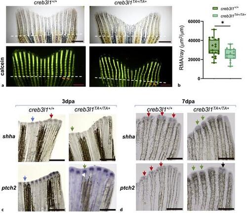
creb3l1TA+/TA+ zebrafish show decreased mineralization and have alterations in expression patterns of Sonic Hedgehog pathway components. a Approximately 50% of the caudal fin was amputated from 6 mpf creb3l1+/+ and creb3l1TA+/TA+ fish. At 9 dpa, the tails were stained with calcein and imaged via brightfield and fluorescence. Representative images are shown. Amputation planes are indicated with dashed lines. Sienna lines in calcein panels mark the width of the 2nd and 3rd rays below the amputation plane. Scale bars = 1 mm. b Fluorescence images analogous to those in (a) were used to measure the fluorescent mineralized area (RMA: total fluorescence distal to amputation plane), which was normalized to the average width of all fin rays below the amputation plane. The creb3l1TA+/TA+ fish exhibit significantly decreased mineralization of the regenerated bone when assessed by an unpaired t-test with Welch’s correction. *p ≤ 0.05. N = 13–19. Each data point represents an individual fish. c, d Approximately 50% of the caudal fin was amputated from 6 mpf creb3l1+/+ and creb3l1TA+/TA+ fish. At 3 dpa (c) and 7 dpa (d), tails were processed for in situ hybridization to detect expression of shha and ptch2 components of the Sonic Hedgehog pathway. Representative images are shown. cshha signal in creb3l1+/+ regenerates is either in a single cap over a regenerating ray (blue arrow) or is already separated into two foci (red arrow). In contrast, in creb3l1TA+/TA+ regenerates, all shha signal is in a single cap over regenerating rays (green arrow). ptch2 signal is in a single cap in both, creb3l1+/+ and creb3l1TA+/TA+ regenerates (blue and green arrow, respectively). d All shha signal in creb3l1+/+ regenerates is separated into two foci (red arrows), but is still within a single cap in creb3l1TA+/TA+ regenerates (green arrows). ptch2 signal in creb3l1+/+ regenerates is separated into two distinct foci (red arrows), but is mostly present in a single diffuse domain in creb3l1TA+/TA+ regenerates (green arrows). Separation of the patch2 signal can be seen in some creb3l1TA+/TA+ regenerates (black arrow). N = 3–7. Scale bars = 1 mm. |